Thursday, December 11, 2014

Be careful! You'll put your eye out.


This Holiday Season, the American Academy of Ophthalmology Urges Caution When Choosing Gifts for a Child.


Though we LOVE the themes of courage, strength, and adventure in many popular films for younger viewers, be aware of your child's desire to appropriate featured weaponry, and avoid buying toys that launch projectiles, such as crossbows and BB guns. 

 
Projectile-shooting toys can cause serious eye injury and vision loss in children. Roughly 1 in 10 children's eye injuries that end up in the ER are caused by toys, according to a 2014 study. Overall, there were an estimated 256,700 toy-related injuries treated in emergency rooms nationwide in 2013, a report by the U.S. Consumer Products Safety Commission found. 


One toy crossbow that shoots darts more than 100 feet away landed on a list of most dangerous toys of 2014 for its potential to cause eye injury. Plastic darts and arrows can scratch the eye, causing corneal abrasions, or in the case of pointed tips can puncture the eye and permanently damage a child’s vision. Injuries from airsoft, BB and paintball guns are quite common and include retinal detachment that can cause vision loss; pooling of blood in the front of the eye (ocular hyphema) that can block vision and increase the risk of glaucoma; and traumatic cataracts, which may require surgery to restore sight.

“People may view toy versions of bows and arrows or guns as harmless, but even foam or plastic projectiles can potentially cause serious damage to a child’s eye if used at close range,” said Jane Edmond, M.D., a clinical spokesperson for the American Academy of Ophthalmology. “With so many other options for gift giving, physicians recommend that parents consider safer alternatives. Nobody wants to end up in the emergency room over the holidays, especially due to an injury caused by a gift.”

Toy safety tips:

 1. Avoid purchasing toys with sharp, protruding or projectile parts such as airsoft guns, BB guns and paintball guns, which can propel foreign objects into the sensitive tissue of the eye.
2. For laser toys, look for labels that include a compliance statement with 21 CFR Subchapter J to ensure the product meets the Code of Federal Regulations requirements for laser products, including power limitations.
3. When giving sports equipment,, provide children with the appropriate protective eyewear with polycarbonate lenses, which are shatterproof.
4. Check labels for age recommendations to be sure to select gifts that are appropriate for a child's age and maturity. Also, keep toys that are made for older children away from younger children.
5. Make sure children have appropriate adult supervision when playing with potentially hazardous toys or games that could cause an eye injury.

References:

Wednesday, November 19, 2014

Contact Care is Critical


Tips for Healthy Contact Habits


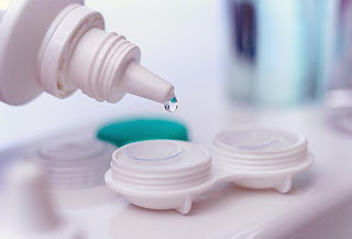
According to the Center for Disease Control, properly caring for your contact lenses has a HUGE impact on your eye health and on the medical industry as a whole. 


 Your Contacts

Replace your lenses as often as the doctor suggests, even if you don't wear them every day. For instance, if you have the disposable kind that are good for one wearing, throw them out after you've worn them once, even if they still feel fresh. When you wear lenses for more days than you should, or when you sleep in lenses that aren't made for overnight wear, you raise your risk of eye infections (Web MD).


Before You Touch Your Lenses
Always wash your hands before you put your lenses in or take them out. Don't lather up with oily or heavily scented soaps. Lenses can cling to wet hands, so dry your hands well with a lint-free towel. If you want to use a moisturizer, wait until after you've put in your lenses. The residue from lotions can stick to them (Web MD). 

Putting in Your Lenses
Start with the same eye each time so you don't mix up the right and left lenses. Use your index finger to slide the lens out of the package or case and into the palm of your hand. Rinse it with the solution recommended by your eye doctor. Place the lens on the tip of your index finger.  Pull your lower lid down with the middle finger of the same hand and hold your upper lid with your other hand. Place the lens directly on the iris of your eye. Gently release your lids, and blink (Web MD).

Removing Your Lenses
To take out soft lenses, pull down your lower lid. Look up or to the side, and gently move your lens to the white of your eye. Using your thumb and index finger, gently pinch the lens and lift it off your eye. For gas-permeable lenses, open your eyes wide and pull the skin near the corner of your eye toward your ear. Bend over your open palm and blink. The lens should pop out into your palm (Web MD).

Cleaning and Storage
There are many ways to clean lenses. A multipurpose solution lets you clean, rinse, disinfect, and store your lenses. Some systems have separate products for cleaning and rinsing. "No-rub" solutions say that rinsing alone will clean the lenses, but research suggests that rubbing cleans better. With hydrogen peroxide solution, you put your lenses in a basket that goes in a cup of solution. Never use this solution to rinse your lenses (Web MD).

Water and Lenses Don't Mix
If you're out of contact lens solution, you might be tempted to rinse your lenses with tap water. Don't do it! Water sometimes has microbes that can cause serious eye infections. Don't even wear contacts in the shower. And never put your lenses into your mouth or use saliva to wet them (Web MD).


The Case Matters, Too
Clean your lens case as carefully as you clean your lenses. You should rinse it at least every night with disinfecting solution. Wipe the case with a tissue and let it air dry to help get rid of lingering bacteria. Replace your case every 3 months or more often (Web MD). Never use water to clean your case.

Your Contact Lens Solution
Don’t “top off” solution. Use only fresh contact lens disinfecting solution in your case—never mix fresh solution with old or used solution. Use only the contact lens solution recommended by your eye doctor.

When Your Lenses Hurt
A lens might feel uncomfortable if there's something on or under it, or if it's inside out. Take the lens out and rinse it with rewetting drops or a non-peroxide solution to remove the dirt or dust. Look at it from the side. If there are marks on the edge and you can read them, the lens is OK. If the shape is a "U," then it's fine. If it's a "V" with the top edges flared out, turn it around, rinse, and place it back in your eye. If that's not better, see your eye doctor (Web MD).

Teens and Contact Lenses
Teenagers can wear lenses as long as they learn to take care of them. Disposable lenses that are worn just 1 day are a good option. There's no cleaning or care involved. Work with your eye doctor to find the best contacts for you. Never try on a friend's lenses. An eye doctor has to fit contact lenses perfectly to your eyes. The doctor can give you samples to make sure the chosen lenses fit well (Web MD).

Makeup Tips for Lens Wearers
Put on soft contact lenses before you apply makeup. Put on gas-permeable lenses after you've put on your makeup. Always take lenses out before you take off your makeup.
Use non-allergenic makeup. Avoid metallic or glittery eye shadows and liners, and lash-lengthening or waterproof mascara. These can irritate or stain your lenses. 
Don't apply eyeliner to your inner rims, between your lashes and your eye.
Replace your eye makeup at least every 3 months (Web MD).

Contact Lenses and Sports
You can wear your lenses for most sports and activities. They rarely move or fall out. Plus, they don't fog up like glasses, and they give you better peripheral vision. If you swim, though, make sure you wear watertight goggles. You can get an eye infection from water, and your lenses can be hard to take out if they get wet. Use saline or rewetting drops to loosen them, and then clean and disinfect them afterward.

Keep Your Glasses
Even after you get contact lenses, you'll probably still wear glasses sometimes. You should keep a pair of up-to-date prescription glasses for when your eyes need a break or if you can’t wear your contacts for some reason. You should also have sunglasses to protect your eyes from damaging UV light. Choose sunglasses that block 99% of UV light and wear them -- especially in the sun, when you're driving, and around snow, water, or sand.

Decorative and Cosmetic Lenses
It might look cool to have cat's eyes for Halloween or change your eye color just for fun. Decorative and cosmetic lenses can be safe, but make sure you get them from an eye doctor. It's actually illegal to sell decorative lenses without a prescription in the U.S. Ill-fitting contact lenses can scratch your eye or cause an eye infection.

Your Eyes and Your Screens
Teens and young adults spend hours in front of computers, televisions, and cell phones. All that screen time can cause computer vision and eye strain problems. We blink less when we're focusing up close like that, so eyes can get dry and tired. To help, try the 20-20-20 rule. Every 20 minutes, take a 20-second break from the screen to look at something 20 feet away (Web MD).    


Visit an Eye Doctor
If you're thinking about contact lenses, see an eye doctor first. Contacts come in different materials, shapes, and strengths that don't match the prescription for your glasses. An optometrist or an ophthalmologist can find the right prescription and fit lenses exactly to your eyes. Even if you want to buy contact lenses online, you'll still need to see an eye doctor first.
(Web MD).


Wednesday, October 15, 2014

Ophthalmology & Art

Ophthalmology and Art:
(Stanford ophthalmologist Michael f. Marmor presents the following research regarding the evolution of Monet’s paintings as impacted by the progression of his cataracts)

Monet, one of the most influential impressionists of the early 20th century, had cataracts that worsened steadily from 1912 to 1922. Slowly progressive age-related cataracts manifest as a yellowing and darkening lens, greatly impacting color perception and visual acuity.

We find striking changes in the style of Monet’s paintings during the period of progressive visual failure. These late paintings show predominant red-orange or green-blue tones that are quite different from the subtle color shading that characterizes Monet’s earlier Impressionistic work. There is nothing in Monet’s correspondence to suggest that he had any intention of mimicking the abstractions and distortions explored by other painters in the early 20th century. It is very difficult to know a century later whether these works appear to us as Monet wanted them to appear. After having his cataracts removed, he destroyed many of his late canvases. Many remain only because they were salvaged by family and friends.

1914-1915: Monet’s visual difficulties were becoming more serious. He wrote that “colors no longer had the same intensity for me . . . reds had begun to look muddy . . . my painting was getting more and more darkened.” He felt that he could no longer distinguish or choose colors well and was “on the one hand trusting solely to the labels on the tubes of paint and, on the other, to force of habit.”

1919-1922: Monet was afraid that he might have to stop painting. He would only paint during certain hours when the lighting was optimal. He was well aware that colors were lost in the yellow blur of his vision.

1923: Monet finally acquiesced to cataract surgery. Though he regained acceptable reading vision, he was acutely aware of changes in color perception in the eye that underwent surgery. He complained vigorously for more than a year that the world appeared either too yellow or too blue. He finally regained confidence in his view of the world in 1924 and worked vigorously to refine the great Water Lily canvases now hanging in Paris, France.

(The following information is from Guy Gugliotta’s article “Simulations of Ailing Artists’ Eyes Yield New Insights on Style,” in the New York Times, 2004)

France in the late 19th and early 20th centuries embraced an astonishing number of important artists who battled serious physical shortcomings — sometimes for decades.

Edgar Degas, known for his paintings of nudes and ballet dancers, suffered retinal disease, probably macular degeneration, for nearly half his life. Degas first noticed eye problems as a national guardsman in the Franco-Prussian War in 1870-71, when he could not aim his rifle because of a blind spot in his right eye. By 1890, his left eye also began to deteriorate. Light dazzled him. He tried to use peripheral vision to compensate for his loss of central vision.
       Retinal disease, unlike cataracts, does not cause major difficulties with color perception. But Degas had blurred vision, affecting his ability to perceive form and line. Dr. Michael Marmor used computer simulations to gauge the problem. The simulations showed that Degas’s draftsmanship became less detailed and the shadowing coarsened as his sight deteriorated. Even so, his work would have looked smoother to him than it actually was.

Mary Cassatt, like Monet, had cataracts. Cataract surgery was possible in the early 1900s, but it did not always work. “I look forward with horror to utter darkness,” Cassatt wrote in 1919, fearing that an operation on her left eye would be “as great a failure as the last one.”

Camille Pissarro had a malfunctioning tear duct, and in his last 15 years, suffered chronic infection of the tear sac in his right eye. Thus, he had difficulty painting outdoors, particularly in winter.        
       Still, “There is a certain element of ‘I’m not going to stop what I want to do,’” said a great-grandson of the artist, Joachim Pissarro, an art historian at Hunter College. “You don’t want to over-analyze the impact.” Indeed, Pissarro’s late cityscapes of Rouen and Paris, regarded as masterpieces, were painted from indoors behind a window to protect his eyes.

For artists with eye problems, it is perhaps surprising that infirmities did not change their styles more radically. A key, some experts said, might be that although artists’ perceptions might be influenced by physical limitations, they are also informed by what the artists know and what they want to do.

“Most of us are into quick snapshots,” said John Elderfield, chief curator of painting and sculpture at the Museum of Modern Art. “But the ability to translate visual memory into a different medium is another thing altogether. Monet had been painting for 50 years when he had cataracts. Of course he painted from memory. He painted from memory all his life.”

*Edited for length
Sources:

Wednesday, October 1, 2014

Diabetes and Your Eyes


If Diabetes impacts you or a loved one, read this wonderfully informative article from DiabeticConnect.


People with diabetes have a higher risk for eye problems. Over time, high blood sugar and high blood pressure levels damage blood vessels throughout the body. This includes the blood vessels that supply the eye.

Scanning Electron Microscope image of retinal blood vessels. 
(source:http://www.gettyimages.com.au/detail/photo/retina-blood-vessels-coloured-scanning-high-res-stock-photography/123773764)

The retina of your eye has tiny, fragile blood vessels on it. When harmed, these blood vessels can swell, weaken and clog. The term used to describe this damage is "diabetic retinopathy," and it’s the leading cause of blindness in adults.

(source: http://peposevision.com/diabetic-retinopathy/)

Having diabetes also ups your risk for cataracts and glaucoma. The longer you have diabetes, the higher your chance of having diabetic-related eye problems.


Signs of diabetic retinopathy include:
1. Blurry, cloudy, or distorted vision. In the early stages of diabetic retinopathy, the blood vessels leak, which can alter your sight. If fluid from the blood vessels leaks into the macula (the inner part of the retina), vision may become blurry.

2. Vision loss or dark area in your vision. Sometimes new blood vessels grow on the surface of the retina after old blood vessels become damaged. The new blood vessels can bleed into the eye and cause vision loss. When diabetic retinopathy is left untreated, scar tissue may build up at the back of the retina, causing the retina to break away from the eye. This is called “retinal detachment”, and it can lead to permanent blindness.

3. Spots floating in your vision (“floaters”). Floaters may be a sign of blood vessel leaks, and they can for days or months. Even if the floater goes away quickly, it’s important to call your doctor before more severe bleeding occurs.

4. Trouble seeing colors. Growing or leaking blood vessels can impair your ability to see colors.

5. Pain, pressure or redness of your eye. Diabetic retinopathy can also lead to macula edema, or swelling of the macula, which can be painful.

6. Double vision. Seeing double may be a sign of cataracts, the clouding of the eye’s lens. Cataracts can also bring on blurred or dimmed vision.

7. Watery eyes, halos, eye aches and headaches. These discomforts may signal glaucoma, which is an increase in fluid pressure inside the eye that pinches the optic nerve. This causes nerve and retinal damage and, eventually, blindness. Other symptoms of glaucoma include blurred vision, pain, and loss of sight.

If you have any of these symptoms or any changes in vision, see your eye doctor at once. Seeking prompt treatment for eye problems may save your vision.

Keep Your Eyes Healthy
Following your diabetes care plan as directed by your doctor can lower your risk of all complications, including diabetic retinopathy. The better you control your blood sugar, the lower your chance of eye problems.

Diabetic retinopathy doesn’t usually cause symptoms in the early stages of the disease, so it’s crucial to get regular eye exams once per year — or as often as your doctor recommends — even if your eyesight seems fine."

Wednesday, September 24, 2014

Ancient Cataract Surgery


When did the first cataract surgery occur?

"That wasn't so bad, now, was it?"

According to the Sushruta Samhita, it was around 800 B.C.E.

This development is contemporary with the Epics of Homer, the first Olympics, India's Iron Age, the Biblical prophet Isaiah, and the adoption of the alphabet by the Greeks. 

THE FIRST OLYMPICS: 776 B.C.E.
THE BIBLICAL PROPHET ISAIAH: 8th CENTURY B.C.E.
THE EPICS OF HOMER: 8TH CENTURY B.C.E.
THE FIRST CATARACT SURGERY IS PERFORMED BY SUSHRUTA, THE "FOUNDING FATHER OF SURGERY": 800 B.C.E.

Here is a translated breakdown of the surgery provided in Duke Elder's Systems of Ophthalmology:

Preoperative care:
1) The patient was recommended to have an oily massage followed by a kind of heat-bath.


Operative technique:

1) The patient was asked to sit on a high stool, with the
surgeon sitting in front of him face to face.


2) He was instructed to look at the surgeon's nose while the operator rested his little finger on the outer eye socket bone, and held a sharp-pointed needle between his thumb, index, and middle

finger.


3) The point entered the anterior chamber, at the

junction of the medial and lateral two-thirds of the outer
portion of the white layer of the eye ball. If a sound
was produced followed by the gushing of watery fluid, then
the surgeon's needle was considered to be in the correct
part of the eye ball, but if this puncture was followed by
bleeding, it meant that it was misplaced. Care was taken
to avoid blood vessels present in that region.


4)The tip of the needle was then made to incise the capsule
of the lens.


5) With the needle in this position, the patient was
asked to blow down the nostril, while closing the opposite
side of the nose.

After this procedure, according to Sushruta, lens
material came out alongside the needle. When the
patient was able to perceive objects, the needle was
removed.


Post-operative care:

A few indigenous medicines (roots and leaves) were applied
to the eye, with a bandage. The patient was then
instructed to lie flat on his back and avoid any movement,
particularly sneezing and coughing. The operated eye was
examined daily until the tenth day. If the whitish mass
appeared again in the pupillary area the same procedure
had to be repeated.


Wednesday, August 13, 2014

Healthy Habits: Cosmetics

Many of us are familiar with the saying, "every old barn needs a little paint." If you embrace this philiosophy, this post is for you.
MISS PIGGY KNOWS A THING OR TWO ABOUT PAPERING HERSELF.
Here are some great tips from the American Academy of Ophthalmology for keeping your eyes healthy as you emphasize their intrinsic beauty:


Throw away eye makeup after three months
Infection-causing bacteria grow easily in creamy or liquid eye makeup. If you develop an eye infection, immediately toss all of your eye makeup.


Never share eye makeup
 AND when sampling makeup in stores, use only fresh applicators and samples that have not been contaminated by multiple users. (The safest choice is to avoid store samples altogether.)


If you tend to be allergic, introduce only one new eye makeup or care product at a time. 
If there is no reaction, add another new product, and so on. If you notice an allergic reaction, find out what the ingredients are and let your doctor know. Avoid products that contain untested or harmful chemicals.


Before applying makeup, be sure your face and eyelids are very clean. 
Always apply makeup outside the lash line, away from the eye, to avoid blocking the oil glands of the upper or lower eyelid. These glands secrete oil that protects the eye’s surface. Never apply makeup while in a moving vehicle.


Do not separate your mascara-clumped lashes with sharp items.


Remove all eye makeup at night before sleeping
 especially mascara that can stick to the lashes. Brush a clean cotton swab along the base of the eyelashes to remove all makeup remnants. If you use eye makeup remover, avoid getting it in your eyes and thoroughly rinse remover off your eyelids.


If you have eye surgery, do not wear makeup around the eye 
until your ophthalmologist tells you it is safe to do so, and then use only fresh, new makeup.


If you tend to have dry eyes, avoid metallic/glitter, powder or other makeup that flakes. 
Flakes can get into the tear film and increase your eyes’ irritation. Glitter eye makeup is a common cause of corneal irritation or infection, especially in contact lens users.


 And here are some more make-up related images for the pure fun of it. 


This information is from eyeSmart, distributing eye health information from the American Academy of Ophthalmology.


Thursday, July 31, 2014

Six Smart Things College Students Should Do for Their Eyes


From the American Academy of Ophthalmology:


American Academy of Ophthalmology offers parents sight-saving advice for teens headed to college


Don't shower or swim in contact lenses. 
Acanthamoeba is a parasite that lives in water and can cause a rare but serious eye infection called Acanthamoeba keratitis. According to the CDC, 85 percent of Acanthamoeba eye infections occur in contact lens wearers, one of the main risks being exposure of lenses to water. To avoid this dangerous infection, do not wear contact lenses in showers, hot tubs or when swimming in lakes or pools. Also, never use water to clean or store contact lenses; only use sterile contact lens disinfecting solution and a clean contact lens case.  


Go outside. 
Scholastically-inclined students spend much of their time studying indoors, which can put them at risk of becoming more nearsighted, or myopic. A 2014 study found that more than 50 percent of college graduates are nearsighted, with eyesight worsening for each year in school. Other research shows that spending more time outdoors can protect vision from getting worse. Head outside when possible.


Wash your hands.
 Conjunctivitis, often called pink eye, spreads fast in schools and dorms. An outbreak struck more than 1,000 Ivy League college students in 2002. Avoid rubbing the eyes and wash hands with soap to avoid catching and spreading pink eye, not to mention other infections.


Give your eyes a break
Nearly 80 percent of engineering and medical school students experienced symptoms such as dry eyes and redness, according to a study of students at one Indian university. To help avoid eye strain, follow the 20-20-20 rule: look at something 20 feet away every 20 minutes for 20 seconds. Because dry eye can also cause painful corneal ulcers, which are open sores on the front part of the eye, blink regularly and fully to keep eyes moist. 


Don't share makeup
Harmless as it may seem, sharing makeup is a surefire way to spread infection such as herpes keratitis among friends. Infection-causing bacteria grow easily in creamy or liquid eye makeup. Stick to your own makeup and throw it away after three months. If you develop an eye infection, immediately toss all of your eye makeup.  

Protect your eyes during the game
Nearly 1 in 18 college athletes will get an eye injury playing sports.[v] Common injuries, like scratches on the eye surface and broken bones near the eye socket, happen most often in high-risk sports such as baseball, basketball and lacrosse. Athletes should consider wearing polycarbonate sports glasses to help keep stray balls and elbows from hitting their eyes


"For many teens just starting college, taking care of their eye health may be the last thing on their minds," said Rebecca Taylor, M.D., comprehensive ophthalmologist and clinical spokesperson for the American Academy of Ophthalmology. "But the fact is that an eye injury or condition can affect their grades and social life, causing days or even a lifetime of poor vision. We hope that parents will remind their kids of these risks before they fly the coop this fall."


For more information on back-to-school eye health, visit the American Academy of Ophthalmology's public education website www.geteyesmart.org.


Wednesday, June 11, 2014

Why Do We Get "Sleep" in Our Eyes?

Ever wonder what those small gold nuggets are doing in the corners of your eyes every morning? You should find this article from the Huffington Post enlightening:

Why do we get "sleep" in our eyes?
       You may call it "sleep," others might call it "crust" or "sand." Some doctors call it "mattering," and a less-commonly used term is "rheum." But no matter the name you use, you know it when you see it -- that white-yellow crumbly gunk on your eyes after you've had some shut-eye.

       The crusty stuff is actually made up of a bunch of different materials, including discarded cells, mucus and debris (including bacteria, bits of oil from the eyelids, and dust), that are collected as the eyelids sweep across the eye, explains Dr. Ivan Schwab, M.D., a professor of opthalmology at the University of California Davis School of Medicine and a member of the American Academy of Ophthalmology.

      Our eyelids close similarly to a zipper, from the cheek side toward the nose. When they do that, they push the tears across the eye, picking up all these different materials along the way. This discharge ultimately collects in the corner of the eyes or the lower lid, and a bit may also collect on the upper lid, he says.
       "It may be a bit like using an exfoliant on your skin: You're cleaning off dead skin or any surface debris you don't want," Schwab explains to HuffPost.

       So is "sleep" clean? Schwab says that it's not sterile -- nothing on your body really is, including the surface of your eye, or even the surface of your skin. That being said, if you use your hands to wipe away eye crust (which you shouldn't be doing in the first place, Schwab says -- a warm, wet washcloth is better for cleaning your eyes) -- it is probably a good idea to wash your hands afterward. 
 
       Normal "sleep" has a slight cream-like tint to it, though it could appear darker if you wear makeup. An active bacterial or viral infection could also increase the amount of eye discharge, and make it more yellow- or green-tinted (at which point, you should see an eye doctor).
Eye discharge that's on the drier side may be indicative of a drier environment, Schwab says. In addition, you might experience more eye discharge if you have an increase in mucus -- allergies are a big culprit.

       "The eye produces mucus in response to allergens in the air," according to Schwab. "Allergies like pollen, they get in the eye [and] they cause the eye to create mucus [that] surrounds the pollen and takes it to the corner" of the eye.

       In addition, contact lens-wearers may experience more eye discharge because they are technically walking around with a foreign body in their eyes all day. If a contact lens fits perfectly, you'll still experience a little excess mucus production -- and if the contact lens fits less than perfectly, or traps debris (such as pollen) beneath the lens, then the eye will produce even more mucus, he says.

       Changes to the amount of eye "sleep" your body is producing could be a sign of an eye infection or other problem. Meanwhile, if you have chronic discharge that hasn't changed (but there's a lot of it), that could reflect something else, such as irritation from dust, or even allergies. If you have so much eye crust along your lashes to the point where your eyes are stuck together in the morning, this could actually be a sign of blepharitis, a disorder where there is a low-grade inflammation of the eyelids.

Article by Amanda L. Chan
in the Healthy Living section
of the Huffington Post

http://www.huffingtonpost.com/2014/05/29/eye-sleep-rheum-sand-crust-mattering_n_5399559.html

Friday, January 10, 2014

New Year, Take Charge of your Health

New Year’s Post

My son, Gideon,  called me recently concerned that his good friend, Brad, in his mid-20s had been diagnosed with diabetic retinopathy. His friend although an insulin-dependant diabetic, didn’t seem to watch his dietary intake very seriously, would eat erratically and drink alcohol like a frat-rat.  He appeared somewhat overweight and rarely exercised. Gideon himself had been in  that rut for sometime and realized although himself not diabetic, should adjust his lifestyle.

Consequently after reading about food choices, adjusting his diet and commencing a vigorous exercise program he lost 20lb and reduced his waste from a 36 to a comfortable 32 inches over a period of 6 months. The enthusiasm was contagious and Brad decided to follow Gideon’s approach.

Within a few month’s Brad lost weight, people commented about his dramatic change in physique, but most importantly his retinopathy was noted to have completely resolved. WIth improved diet, eating habits and regular exercise Brad’s diabetic control dramatically improved.

Brad had picked up a copy of Richard Bernstein, M.D.’s book entitled: Diabetic Solution.. Diabetics all know that their greatest struggle is controlling their blood sugar levels. In this book, Dr. Bernstein details a ‘solution’ to obesity, fatigue, and many of the health risks associated with diabetes by following a structured diet controling protein, fat, and complex carbohydrate intake. Dr. Bernstein himself has had insulin-dependant diabetes for over sixty years since. After years of fighting with this metabolic disorder, he entered medical school at age 45 determined to discover a way to keep his blood-sugar levels normal. It was primarily in adjusting food intake that he saw dramatic improvement in diabetic control that he could achieve for both himself and his patients.  Since opening his own practice in 1985 Bernstein has helped hundreds of patients improve their diabetic control with dietary restraint.  

As an ophthalmologist, I see a tremendous amount of ocular complications including retinal hemorrhage, retinal swelling and detachment in patients with uncontrolled diabetes. Poor control can also cause cataracts and increase risk of severe infections. There is also a higher chance of developing glaucoma which can cause blindness.

By managing blood sugar levels through a modified appropriate to diet and exercise, many of these problems can be avoided or even reversed. Look for more updates to my blog including fun and healthy recipes that will change your life and your health.

Dr. Bernstein’s Website